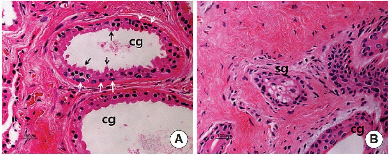
Fig. 1.

Hematoxylin and eosin staining in the ceruminous gland (A) and sebaceous gland (B). The ceruminous gland is a simple coiled tubular gland. The gland is lined by a single layer of secretory cells resting on myoepithelial cells (white arrows). The glandular epithelium is cuboidal or columnar and has protrusions (black arrows) extending to the lumen of the tubule. cg, ceruminous gland; sg, sebaceous gland (H&E,×200).
