
Fig. 3.
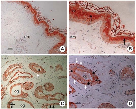
Immunohistochemical labelling of calcitonin gene-related peptide (CGRP) using an avidin-biotin peroxidase complex method in the external auditory canal skin. (A) CGRP positive staining is prominently noted in the suprabasal portion of epithelium (×100). (B) Positive stained cells contain granular and prickle cell layers, in the epithelium (black arrows) (×200). (C, D) Reactions for CGRP were generally moderate in the secretory cells (black arrows), with their myoepithelial cells (white arrows) showing a stronger reaction. The CGRP staining in the sebaceous gland (sg) is somewhat weak, compared to other regions. ep, epithelium; dm, dermis; cg, ceruminous gland. (C, ×100; D, ×200).