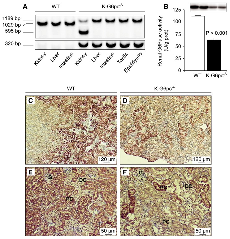
Figure 1. Partial deletion of renal G6pc in K-G6pc−/− mice.
(A) Kidney-specific excision of G6pc exon 3. Genomic PCR analysis of purified DNA from the kidney, liver, intestine, testis and epididymis revealed products of 1189, 1029 and 595 bp corresponding to the floxed, wild-type and excised G6pc alleles, respectively. Specific primers were used to amplify a 320 bp Cre fragment in K-G6pc−/− mice. (B) Renal G6Pase activity in WT (white bar) and K-G6pc−/− (black bar) mice after 6 h of fasting. The results are expressed as the mean ± SEM (n = 12 mice per group). The top panel shows western-blot analysis of G6PC in WT and K-G6pc−/− kidney homogenates. (C–D) Immunohistochemical analyses of G6PC in the kidneys of WT mice (C and E) and K-G6pc−/− mice (D and F). G: glomeruli, DC: distal convoluted tubules, PC: proximal convoluted tubules.

