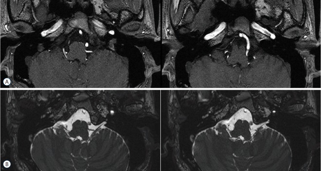
Fig. 1.
Magnetic resonance image of the left posterior inferior cerebellar artery in contact with the root entry zone of the left glossopharyngeal nerve. A: Three-dimensional time-of-flight magnetic resonance angiography (3D-TOF-MRA). B: Three-dimensional fast imaging employing steady-state acquisition (3D-FIESTA).

