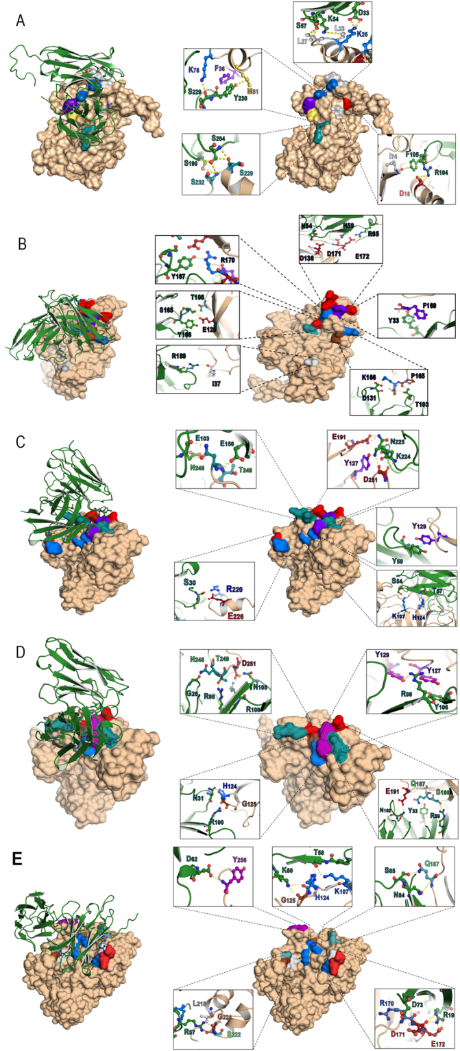
Figure 9.

Computerized interaction of modeled-NS5A and HuscFvs and residues that were predicted to form contact interface between them. Left side of Panels A-E shows interactions of NS5A (beige) and respective HuscFvs (green). Right side of Panels A-E shows contact residues between NS5A and HuscFv5, HuscFv9, HuscFv16, HuscFv19 and HuscFv99, respectively. The NS5A amino acids are colored according to CINEMA color scheme: polar negative D and E are red; polar positive H, K, and R are blue; polar neutral S, T, N, and Q are green; non-polar aromatic F and Y are purple/magenta; non-polar aliphatic A, V, L, I, and M are white (grey in this study as the background is white); and P and G are brown. The π-π interaction is shown as dotted green line; H bond/salt-bridge is shown as dotted yellow line.
