Figure 4.
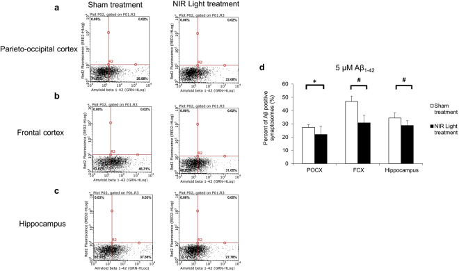
Flow cytometry analysis of ex vivo synaptic binding of 5 μM Aβ oligomers. Representative flow cytometry analysis of the 5 μM Aβ oligomer binding in synaptosomes isolated from (a) parieto-occipital cortex (POCX) (b) frontal cortex (FCX) and (c) hippocampus in NIR light treated and sham treated wild type mice. (d) The results indicate a significant reduction in the percentage of Aβ positive synaptosomes in the three brain regions POCX, FCX and hippocampus of NIR light treated mice (black) compared to sham treated mice (white). (n = 8; per group). Statistical significance was determined by Student’s two tailed t-test analysis. Error bars represent standard deviation. *p < 0.05; #p < 0.01.

