Figure 1.
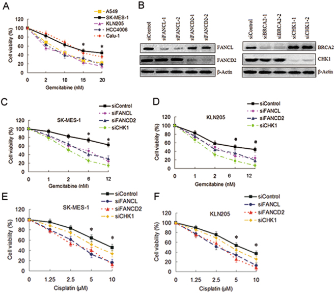
Depletions of the FA pathway factors increased the sensitivity of gemcitabine to SK-MES-1 cells. (A) A549, SK-MES-1, KLN205, HCC4006 and Calu-1 cell lines growing in 96-well plates were treated with gemcitabine at the indicated dose for 4 h. The CCK-8 assay was used to determine cell viability (*SK-MES-1 or Calu-1 as compared with A549, KLN205, and HCC4006 respectively, P < 0.05). (B) Western blot of FANCL-, FANCD2-, BRCA2- and CHK1-depleted SK-MES-1 cells to verify the efficiency of the siRNA transfections. β-actin was used as loading control. Uncropped images are presented in Supplementary Figure S5. (C) SK-MES-1 and (D) KLN205 cells growing in 96-well plates were transfected with various siRNAs as indicated. Cell viability was detected by the CCK-8 assay following gemcitabine treatment for 4 h (*siContrl as compared with siFANCL and siFANCD2, P < 0.005, compared with siCHK1, P < 0.001; #siControl as compared with siFANCL and siFANCD2, P < 0.05, compared with siCHK1, P < 0.005). (E) SK-MES-1 and (F) KLN205 cells growing 96-well plates were transfected with various siRNA as indicated. Cell survival was measured by the CCK-8 assay following cisplatin treatment (siControl as compared with siFANCL and siFANCD2, *P < 0.01).

