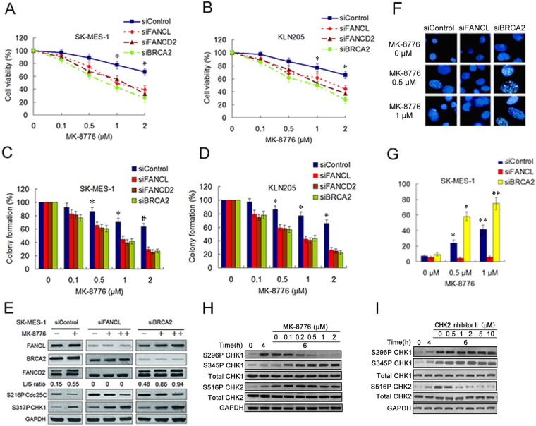
Figure 2.
Depletions of the FA pathway factors sensitized LSC cells to MK-8776. (A,B) The viability analysis were performed using the CCK-8 assay in SK-MES-1 and KLN205 cell lines depleted of the FA pathway factors after MK-8776 treatment for 6 h. (siControl as compared to siFANCL, siFANCD2 and siBRCA2, *P < 0.05; # P < 0.01). (C,D) The two LSC cell lines were treated with MK-8776 following transfection with various siRNA as indicated. The cells were stained by crystal violet and total colonies were counted after two weeks. Colony numbers of control-treated cells were set as 100%. (siControl as compared to siFANCL, siFANCD2 and siBRCA2, * P < 0.01; # P < 0.005). (E) Western blotting detecting monoubiquitination of FANCD2 and phosphorylation of Cdc25C (S216) and CHK1 (S317) in SK-MES-1 cells transfected with siControl, siFANCD2 and siBRCA2. The cells were either treated with MK-8776 (0.5 μM+ , 1 μM++) or DMSO control for 6 h. GAPDH was used as loading control. Uncropped images are presented in Supplementary Figure S6. (F) SK-MES-1 cells were treated with MK-8776 at the indicated doses for 6 h following transfection with siFANCL or siBRCA2, and fixed and immunostained using anti-FANCD2 antibody. (G) The percentage of cells with >20 FANCD2 foci was quantified using Spot Advanced RT software (the cells treated with 0.5 μM MK-8776 as compared to the cells treated with 0 μM MK-8776, * P < 0.05, # P < 0.01; the cells treated with 1 μM MK-8776 compared to the cells treated with 0 μM MK-8776, ** P < 0.01, ## P < 0.001). (H and I) SK-MES-1 cells were treated with 5 nM gemcitabine for 4 hours. The drug was then replaced with MK-8776 or CHK2 inhibitor II at the indicated doses and the cells harvested after an additional 6 hours. Cell lysates were analyzed by Western blotting for the indicated protein.

