Figure 3.
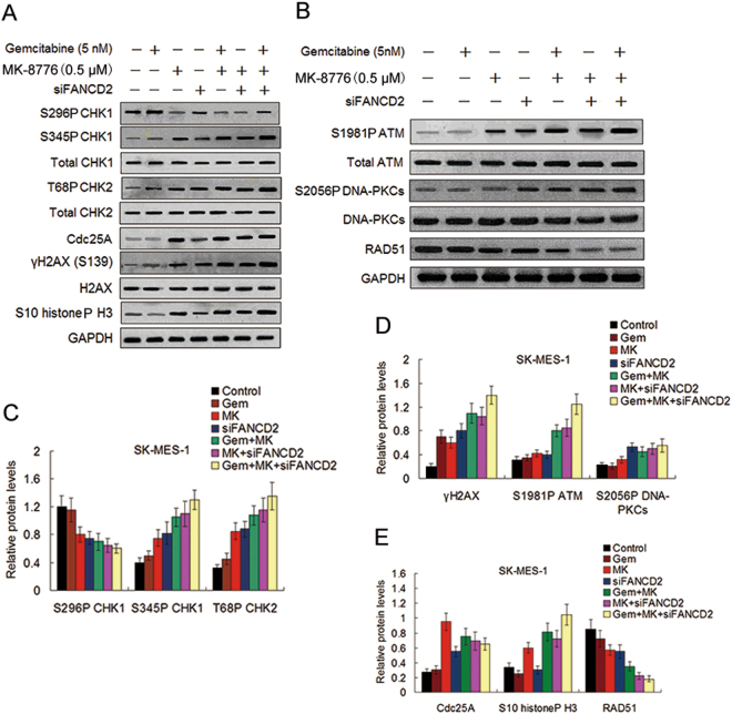
Effect of MK-8776 and FANCD2 knockdown on gemcitabine-induced phosphorylation and expression of proteins involved in the cell DNA damage checkpoint pathways. (A,B) Before and after siFANCD2 transfection, the SK-MES-1cells were treated with gemcitabine for 4 h, or MK-8776 for 6 h, or co-treated with gemcitabine and MK-8776 for 4 h and then with MK-8776 alone for another 6 h after removal of gemcitabine. Cell lysates were analyzed by Western blotting for the indicated proteins as described in the materials and methods, GAPDH was used as a loading control. Uncropped images are presented in Supplementary Figure S7A and B. (C) The intensity of S296P CHK1, S345P CHK1, T68P CHK2, and (D) γH2AX, S1981P ATM, and S2056P DNA-PKCs bands shown in (A,B) was quantified by densitometry and normalized against that of their nonphosphorylated bands. (E) The intensity of Cdc25A, S10 histoneP H3, and RAD51 bands was quantified by densitometry and normalized against that of the GAPDH bands.

