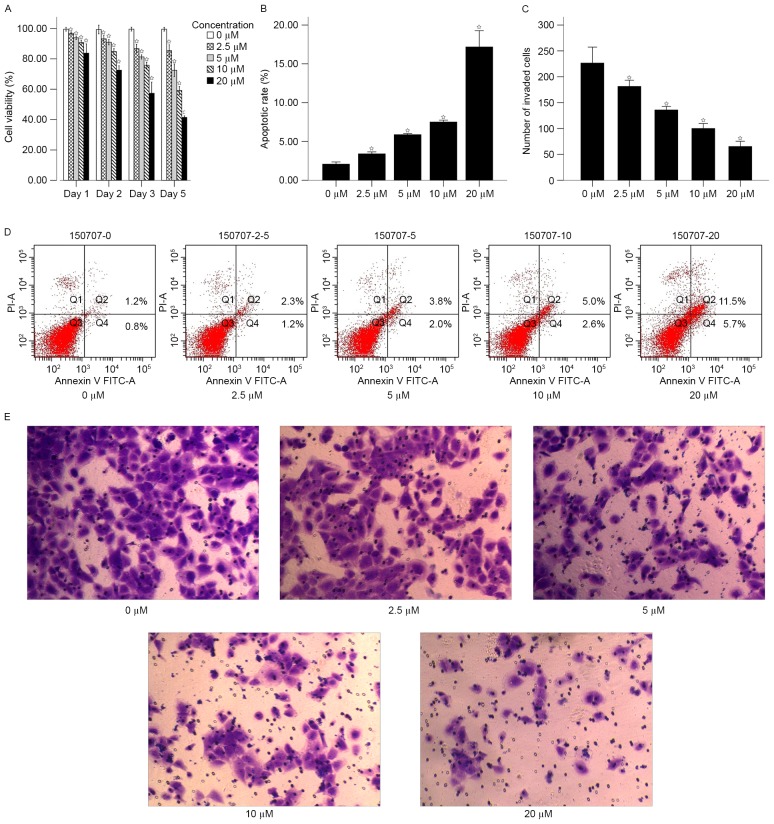
Figure 3.
Determining the effects of re-expression of EN2 and 5-Aza-dc treatment on the apoptotic and invasive capacities of 786-O cells. (A) Effects of 5-Aza-dc on the proliferation of 786-O cells analyzed using MTT assay. Cell viability was assessed at 1, 2, 3 and 5 days post-treatment with various concentrations of 5-Aza-dc (0, 2.5, 5. 10 and 20 µM). (B) The effect of 5-Aza-dc treatment on apoptosis of 786-O cells as analyzed by fluorescence activated cell sorting assay. The cells were treated with various concentrations of 5-Aza-dc for 5 days. (C) Effect of 5-Aza-dc treatment on invasive capacity of 786-O cells as analyzed by Transwell assay. The cells were treated with various concentrations of 5-Aza-dc for 5 days. (D) The apoptosis assay was assessed in different groups that treated with various concentrations of 5-Aza-dc for 5 days. (E) Results of different groups transwell assay that treated with various concentrations of 5-Aza-dc for 5 days. Original magnification, ×200. *P<0.05 vs. control (0 µM group). EN2, engrailed-2; 5-Aza-dc, 5-aza-2-deoxycytidine; FITC, fluorescein isothiocyanate; PI, propidium iodide.

