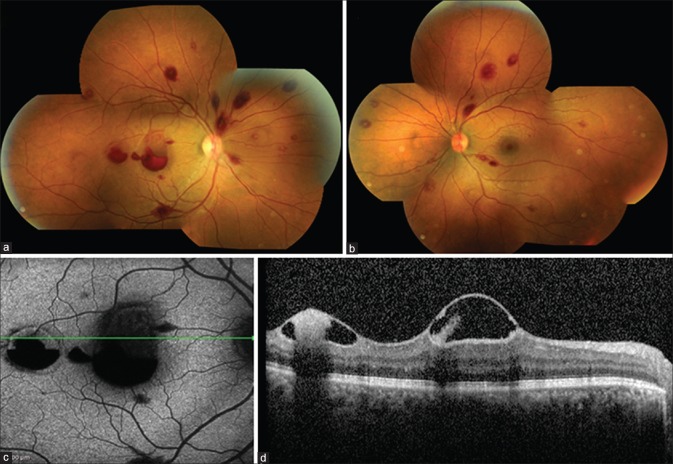
Figure 1.
At presentation, fundus photograph (a and b) of the right eye and the left eye showing multiple Roth spots. The right eye had preretinal hemorrhage over the fovea and temporally. Fundus autofluorescence of the right eye (c) showing hypoautofluorescence corresponding to the hemorrhages. Optical coherence tomography of the right eye (d) showing elevated hyperreflective internal limiting membrane over the fovea and temporally with subinternal limiting membrane hyperreflective deposits within a hyporeflective cavity due to plasma erythrocyte separation

