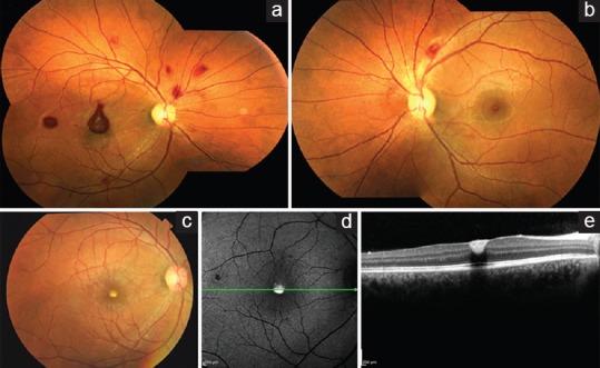
Figure 2.

Fundus photograph of the right eye (a) and the left eye (b) at 2 weeks showing decrease in hemorrhages. Fundus photograph of the right eye at 6 weeks (c) showing complete resolution of hemorrhages and a yellowish spot at the macula. Corresponding fundus autofluorescence (d) showing hyperautofluorescence and optical coherence tomography (e) showing flattened foveal contour with subinternal limiting membrane hyperreflective deposits
