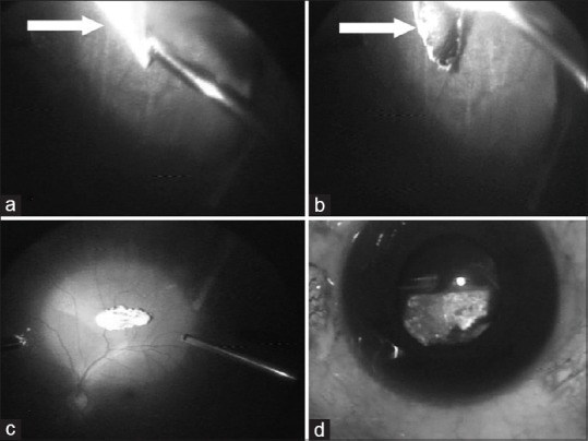
Figure 3.

(a) Intraoperative photograph showing the encapsulated intraocular foreign body at 6 o’clock position near pars plana. (b) The encapsulation being cut with a cutter to free the foreign body. (c) Intraocular foreign body dislodged into the vitreous cavity. (d) Intraocular foreign body being exteriorized from the eye
