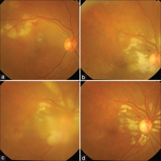
Figure 1.

A photo collage demonstrating worsening of the fundus picture in the right eye with oral antiviral agents and steroids, followed by resolution with oral doxycycline. (a) Cotton wool spots suggestive of retinitis patches in the superotemporal quadrant along the vessel distribution. (b) Increase in the retinitis patches after a course of oral acyclovir, this time in a peripapillary distribution. Hard exudates in the macular area owing to resolution of the macular edema. Superficial hemorrhage superotemporal to disc. (c) Further increase in the retinitis patches along the vessels radiating superior to disc and increase in vitritis (after a week of oral steroids). Steroids were discontinued at this point. (d) Dramatic resolution with reduction in the retinitis patches and vitritis after 1 week of oral doxycycline therapy
