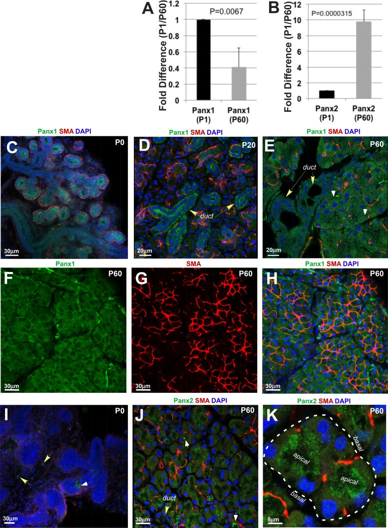
Figure 1.
Panx1 and 2 expression in mouse LG. (A) Panx1 expression slightly decreases in the LG of adult (postnatal day 60 [P60]) mice compared with the LG obtained from mice at postnatal day 1 (P1). (B) Expression of Panx2 increases in adult LGs compared with LGs at P1. In A and B, histograms represent the normalized expression of Panx1 and 2 to GAPDH assessed by qRT-PCR in three independent experiments. (C–H) Panx1 expression in mouse LGs at P0 (C) during postnatal (D: P20) development and adulthood (E–H). At P and P20, Panx1 (green) is highly expressed in the developing LG ducts (C, D). Panx1 expression in the developing acini is low (D). Myoepithelial cells (red) are localized around acini, nuclei labeled with 4′,6-Diamidine-2-phenylindole (DAPI) (blue). (E, F, H) Panx1 expression in adult LG (P60). (F–H) MECs have low level of Panx1 expression. Low level of Panx1 expression in MEC is confirmed in nonmerged images (F, Panx1 expression; green; and G, SMA [marker of MECs] expression, red). (I) Low level of Panx2 expression in the epithelial components of the LG at P. Panx2 was only detected in blood vessels (white arrow) and some mesenchymal cells (yellow arrows) At P60, Panx2 (J, K) is expressed in the acinar cells. High levels of Panx2 protein is found in blood vessels (J, white arrows) and apical parts of acinar cells (K, apical; basal parts of acinar cells are labeled with puncture line). Myoepithelial cells (red). Nuclei are labeled with DAPI (blue).

