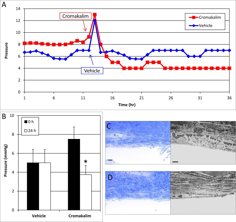
Figure 2.
Effect of cromakalim on intraocular pressure in human anterior segment perfusion culture following removal of the trabecular meshwork and parts of Schlemm's canal. (A) Representative graph of cromakalim treatment showing IOP reduction in human anterior segments with trabecular meshwork and parts of Schlemm's canal removed. (B) Treatment with cromakalim significantly lowered pressure from 7.5 ± 1.3 to 3.8 ± 1.0 mm Hg within 24 hours (P = 0.004), while pressure in the vehicle-treated contralateral eyes remained unchanged at 5.4 ± 1.4 mm Hg (n = 4). (C) Toluidine blue–stained thin sections (100 μm) and transmission electron micrographs of vehicle and (D) cromakalim-treated eyes show complete removal of the trabecular meshwork and the inner wall of Schlemm's canal. Only parts of the outer wall of Schlemm's canal can be seen. The remaining cells and tissues of the distal outflow pathway appear healthy and viable in both control and treated eyes. Scale bar: 20 μm for toluidine blue sections; 5 μm for transmission electron micrographs.

