Fig. 1.

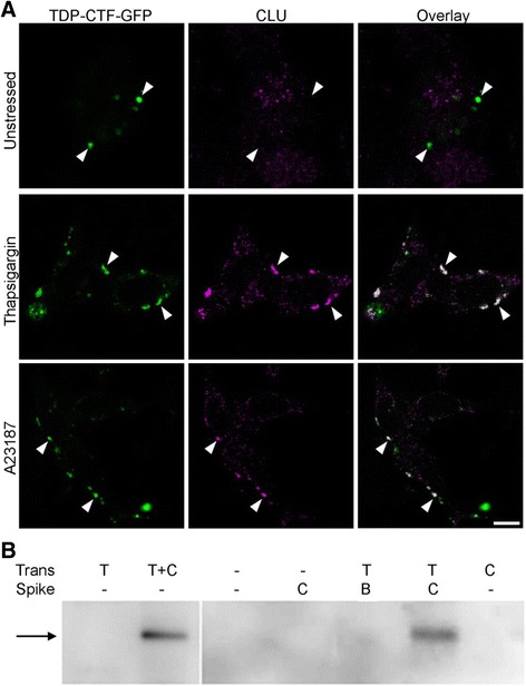
CLU co-localizes with cytoplasmic TDP-43 inclusions in ER stressed cells and co-precipitates with TDP-43-GFP. a N2a cells transfected to express TDP-43CTF-GFP and human CLU (hCLU) were treated as indicated, fixed/permeabilized, then immunostained for hCLU. White arrowheads indicate the positions of inclusions. Overlay panels (right): where there is no colocalization, inclusions appear green; colocalization of TDPCTF-GFP and CLU appears as white. Scale bar is 10 μm; images shown are representative of many. Manders overlap coefficients (TDP-43CTF-GFP and CLU), calculated using at least 20 cells for each treatment, were: untreated, 0.13 +/− 0.14; Tg, 0.62 +/− 0.07; A23187, 0.52 +/− 0.09 (mean +/− SD). See also Additional file 1: Figure S3. b TDP-43 immunoblot showing that TDP-43M337V-GFP co-precipitated with hCLU from lysates of co-transfected N2a cells and also when exogenous purified hCLU (but not BSA) was added (“spiked”) to lysate prepared from N2a cells transfected to express only TDP-43M337V. The black arrow indicates the expected size of TDP-43M337V-GFP. Key above image indicates which protein(s) cells were transfected to express (Trans), and the protein added to the lysate (Spike). The result shown is representative of two independent experiments
