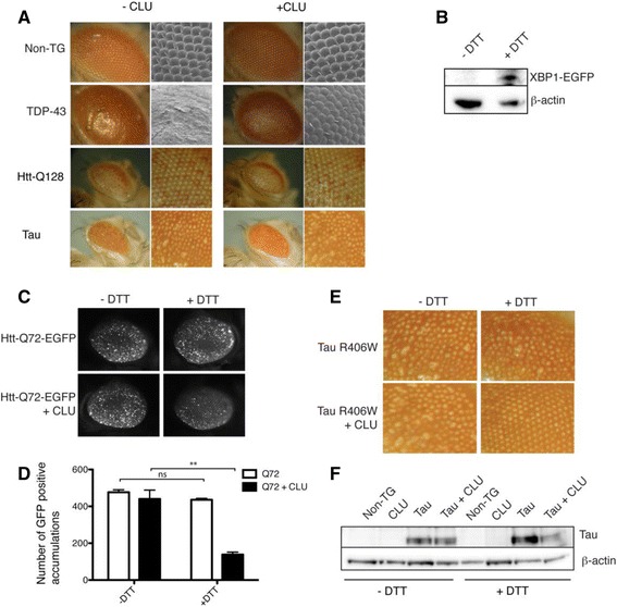
Fig. 6.

CLU provides ER stress-dependent protection against proteotoxicity. a Light and scanning electron micrographs demonstrating the effects of expression of TDP-43, Htt-Q128 and tau R406W (+/− CLU) in the photoreceptor neurons of adult Drosophila. Light micrographs (left) of Drosophila eyes collected using a 7X objective, electron micrographs (right) taken at 200X magnification. For Htt-Q128 and tau R406W, the images shown on the right are optical zooms of the corresponding images on the left. All images are representative of many experiments. b Western blot of whole non-transgenic Drosophila head lysates prepared from Drosophila fed normal food (−DTT) or food supplemented with DTT (+DTT); detection of XBP1-EGFP indicates activation of the UPR (β-actin was used as a loading control). c Fluorescence micrograph images (collected using a 7X objective) of eyes on Drosophila fed with food +/− DTT (or not), and expressing Htt-Q72-EGFP +/− CLU. d Quantification of the number of individual EGFP accumulations per eye, using images such as those shown in (c) and ImageJ (particle analyser program); **p = 0.0037, n = 9, Student’s t-test. Results shown are representative of several independent experiments. e Light micrographs (collected using a 7X objective) showing the eye phenotype in adult Drosophila fed normal food (−DTT) or food supplemented with DTT (+DTT) resulting from expression of R406W tau +/− CLU in the photoreceptor neurons. f Western blot of Drosophila head homogenates prepared from non-transgenic (Non-TG) Drosophila or Drosophila described in (e)
