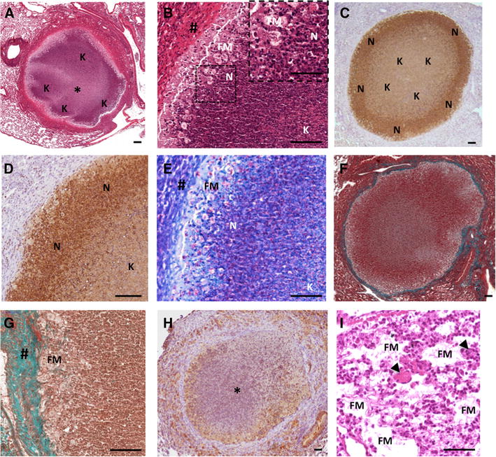
Figure 4.

The C3HeB/FeJ Type I lesions recapitulates the features of the stage III/IV cattle granuloma lesions. A H&E staining of a Type I granuloma showing acellular caseum (*) surrounded by typical neutrophil-derived karyorrhectic debris areas (“K”). B Higher magnification of A showing a characteristic dark stained neutrophil dense area (“N” & Zoom) next to the foamy macrophages ring (FM) below the fibrous capsule (#). C, D Immunohistochemical staining with Ab against Ly-6G (clone 1A8) showing the high density of neutrophil within the subscapular area (“N”) and neutrophil-derived karyorrhectic debris within the core of the lesion area (“K”). E Ziehl staining of the same area as B showing peripheral cellular bacilli within foamy macrophages (“FM”) and neutrophil (“N”) and central acellular bacilli within the karyorrhectic debris (“K”). F, G Masson-Goldner’s Trichrome staining showing the collagen capsule all around a Type I lesions (F). G Higher magnification of F showing the collagen staining among the fibroblast in top of a rime of foamy macrophages. # fibrous capsule, FM foamy macrophages. H Pimonidazole staining showing characteristic hypoxia within central necrotic granuloma (*) and its periphery. I H&E staining, high magnification of foamy macrophages (“FM”) and epitheliod cell dense area containing multinucleated Langhans giant cells (black arrows). Scale bars: 50 μm (B-Zoom, I); 100 μm (A–H).
