
Figure 1.
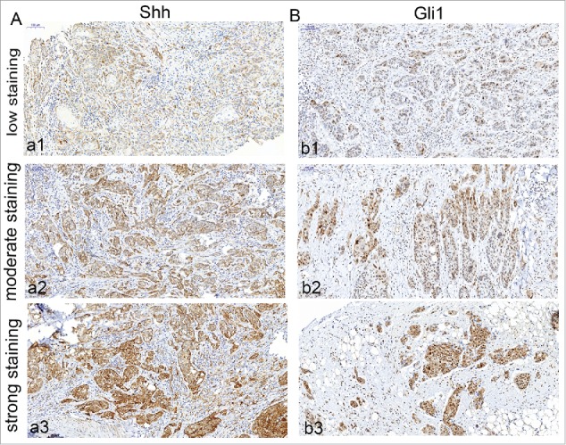
Immunohistochemical detection of Shh, Gli1 in OSCC specimens. A, cytoplasm expression of OSCC specimen for Shh (a1, low staining; a2, moderate staining; a3, strong staining); B, nuclear and cytoplasm expression of OSCC specimen for Gli1 (b1, low staining; b2, moderate staining; b3, strong staining). Bars indicate 100 um.