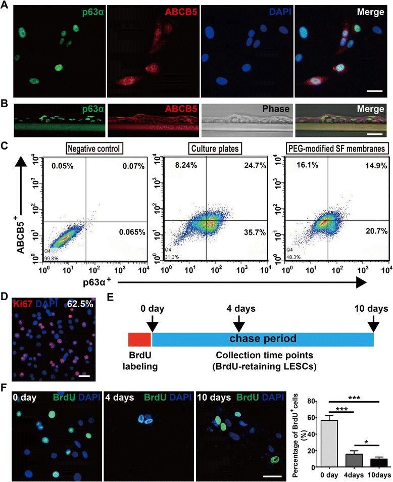
Fig. 5.

Rabbit LESCs cultured on PEG-modified SF membranes maintain characteristics of stem cells. a, b LESCs from the tissue explant cultures expressed p63α and ABCB5 on PEG-modified SF membranes. c Flow cytometric analysis for p63α and ABCB5 of LESCs cultured on culture plates and PEG-modified SF membranes. d LESCs cultured on PEG-modified SF membranes maintained high proliferative capacity (Ki67 staining). e Schematic summary of the experimental design for BrdU chase experiments. LESCs labeled by BrdU for 1 day, and chased in BrdU-free DMEM/F12 medium for 10 days. f Specific staining of BrdU-retaining LESCs cultured on PEG-modified SF membranes at 0, 4, and 10 days. Percentage of BrdU-retaining LESCs was quantified. Data was shown as mean ± SD from three independent experiments. One-way ANOVA analysis: *P < 0.05; ***P < 0.001. Scale bar, 25 μm. ABCB5 sub-family B, member 5, BrdU 5-bromo-2′-deoxyuridine, DAPI 4′,6-diamidino-2-phenylindole, LESC limbal epithelial stem cell, PEG poly(ethylene glycol), SF silk fibroin
