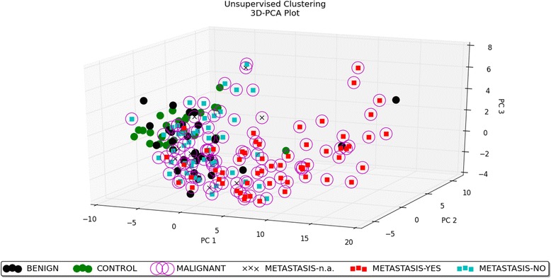
Fig. 1.

3D–PCA plot − unsupervised analysis. PCA performed on all the samples and on the top 50 microRNAs with the highest standard deviation. The normalized log-transformed Hy3 values were used for the analysis. The features were shifted to be zero centred, (i.e. the mean value across samples was shifted to 0) and scaled to have unit variance (i.e. the variance across samples was scaled to 1) before the analysis. PCA plot reveals the distinct sample clusters for metastatic tumours, non-metastatic tumours and the control group
