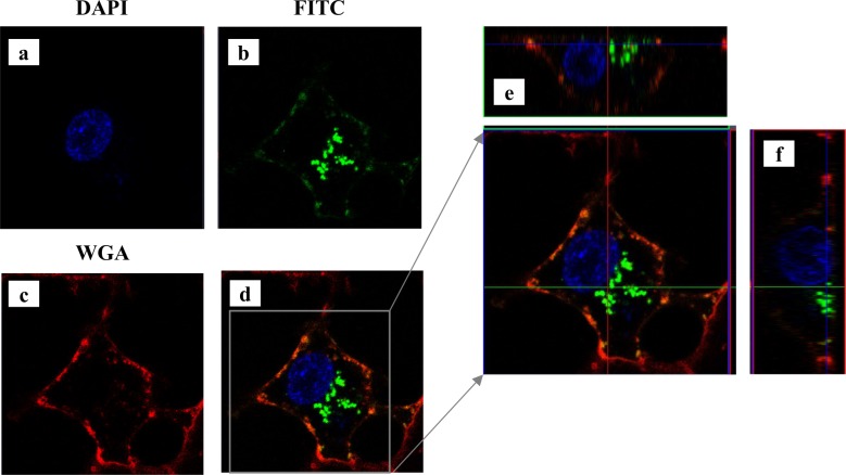
Fig 5. Endocytosis FITC-labeled galectin-7 viewed by confocal microscopy.
Confocal microscopy of MDA-MB-231 cells treated with FITC-rhGal-7 during 25 minutes. Staining with DAPI (a) and WGA (c), which target the nucleus and plasma membrane respectively, are shown. In (b), staining of FITC-gal-7. (e) and (f) show cross sections of the cell.

