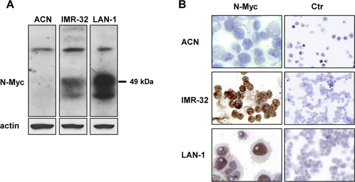
Fig 1. Expression levels and localization of N-Myc protein in NB cell lines.
(A) Protein lysates from ACN, IMR-32, and LAN-1 cells were subjected to Western blot and probed with anti-N-Myc antibody. The blot was reprobed with anti-actin antibody as loading control. (B) ACN, IMR-32, and LAN-1 cells were cytospinned on Polysine™ slides and fixed. N-Myc protein nuclear localization was visualized by immunocytochemistry with anti-N-Myc antibody (60x magnification). Ctr = negative controls (20x magnification).

