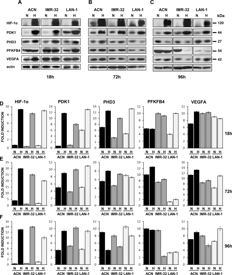
Fig 2. Hypoxia modulates expression of NB-hypo selected genes in NB cell lines.
Protein lysates from ACN, IMR-32, and LAN-1 cells, cultured in normoxic (N) or in hypoxic (H) conditions for (A) 18, (B) 72, or (C) 96 hr, were subjected to Western blot and probed with anti-HIF-1α, anti-PDK1, anti-PHD3, anti-PFKFB4, and anti-VEGFA antibodies. The blot was reprobed with anti-actin antibody as loading control. Levels of HIF-1α, PDK1, PHD3, PFKFB4, and VEGFA in NB cells cultured in normoxic or in hypoxic conditions for (D) 18, (E) 72, or (F) 96 hr were quantified by densitometry and normalized to the content of the loading control protein. The optical density of the scanned film was measured with Quantity One v. 2–3 Image software (Versa Doc, Bio-Rad, Hercules, CA, USA). Results represent the mean values ± S.D. from three independent experiments.

