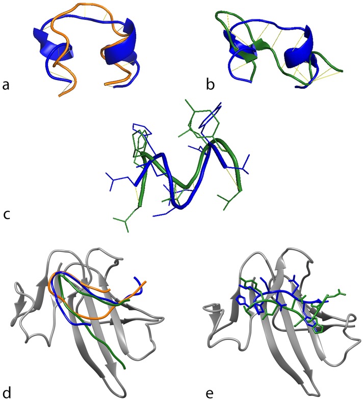
Fig 4. Alignment and docking of human, bovine, and canine (short) Link-N variants.
Schematic of the predicted molecular model and alignment of human (blue) and bovine (orange) Link-N (a) and human (blue) and canine (green) Link-N (b). (c) Schematic of the predicted molecular model and alignment of human (blue) and canine/bovine (green) short Link-N. (d) Docking of human (blue), bovine (orange) and canine (green) Link-N to the extracellular domain of BMPRII. (e) Docking of human (blue) and canine/bovine (green) short Link-N to the extracellular domain of BMPRII. Models represent best-fit predictions for their interaction.

