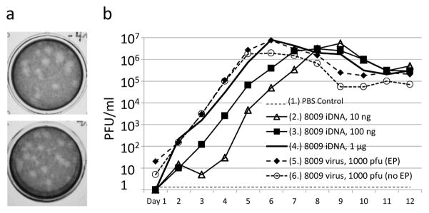
Fig. 3.
Detection of JEV virus in the medium from Vero cells transfected with pMG8009 iDNA plasmid. (a) Plaque assay in BHK cells. Upper panel, plaque assay of growth medium from virus-infected Vero cells (no electroporation), sample taken on day 7 post-infection with 1000 PFU (sample #6 on Fig. 3b). Lower panel, plaque assay of growth medium from pMG8009-transfected Vero cells (after electroporation), sample taken on day 7 post-transfection with 10 ng of DNA (sample #2 on Fig. 3b). (b) Growth curves of JEV virus in the medium of Vero cells transfected with pMG8009 iDNA (samples 2, 3, 4) or infected with pMG8009-derived virus (samples 5 and 6). Samples 5 and 6 show infection with 103 PFU of pMG8009-derived vaccine virus of electroporated and non-electroporated Vero cells, respectively.

