
Figure 4. Action of CK1δ within the purified nuclear PER complex.
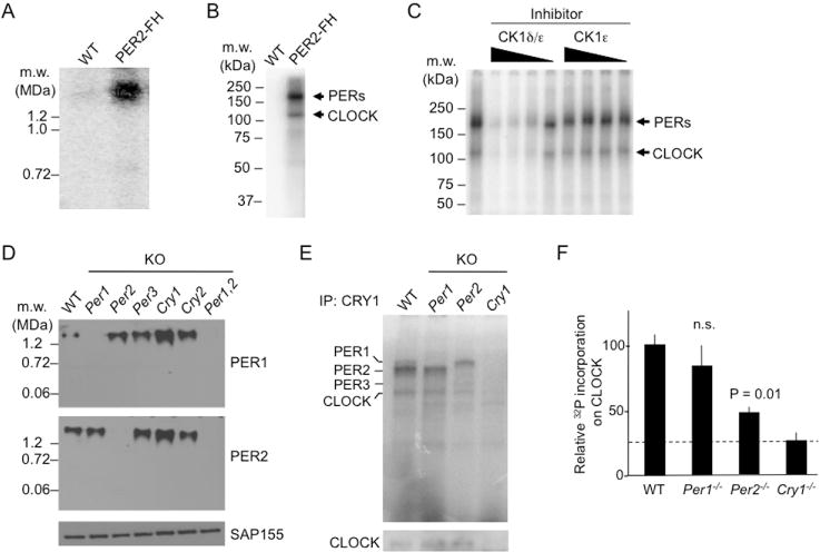
(A) Protein kinase activity of nuclear PER complex. BN-PAGE autoradiogram showing control sample (WT) or affinity-purified nuclear PER complex (PER2-FH) after incubation with γ-32P-ATP (25°C, 1 h).
(B) SDS-PAGE autoradiogram of material analyzed in (A). See Figure S5.
(C) CK1δ in purified PER complex phosphorylates PERs and CLOCK. SDS-PAGE autoradiogram as in (B). Leftmost lane, no inhibitor. Remaining lanes, decreasing concentrations (625, 125, 25, or 5 nM, represented by wedges) of PF-670462, a CK1δ/ε inhibitor (CK1δ/ε), or PF-4800567, a CK1ε-selective inhibitor (CK1ε).
(D) Top and Middle, BN-APAGE immunoblots of mouse liver nuclear extracts (CT18) from wildtype (WT) or single mutants null for the circadian clock genes indicated at the top (KO) probed for proteins indicated at right. Bottom, SDS-PAGE SAP155 immunoblot loading control.
(E) Top, SDS-PAGE autoradiogram of liver nuclear extracts (CT18) from genotypes indicated at top after immunoprecipitation with anti-CRY1 antibody and incubation with γ-32P-ATP (25°C, 10 min; linear range for wildtype). Bottom, SDS-PAGE immunoblot for CLOCK served as loading control for PER complexes.
(F) Phosphor-imaging quantification of labeled CLOCK bands from (E) after normalization to CLOCK immunoblot signals from (E). Shown are mean ± SEM; t-test (two-tailed). Dashed line, background.