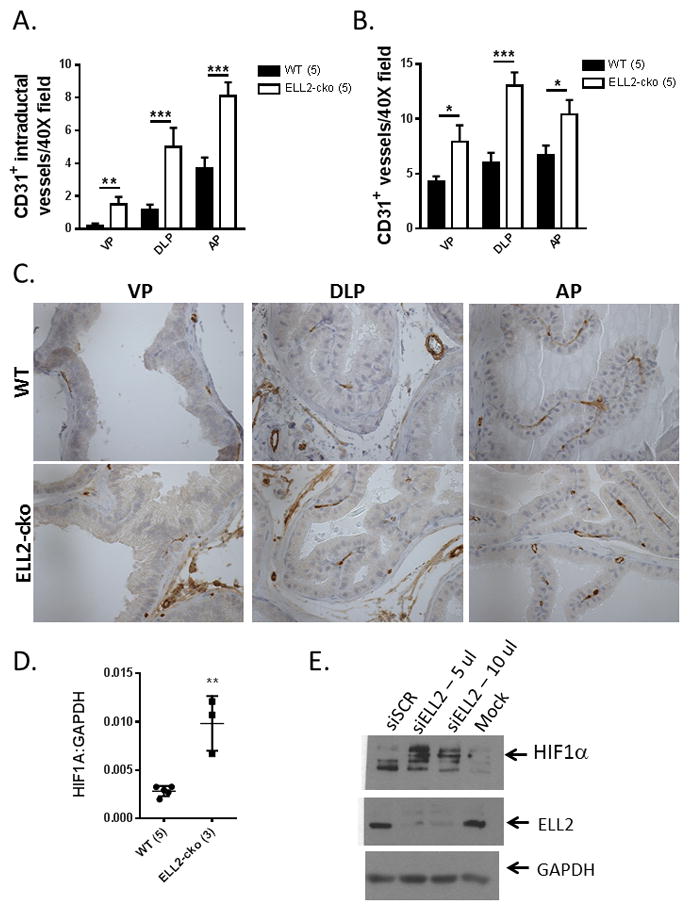
Figure 4.

CD31-positive microvessel density in Ell2-cko mice at age 17–20 mos. A. Quantification of CD31-positive intraductal microvessels in Ell2-cko mice vs wild-type (WT) controls. B. Quantification of total CD31-positive microvessels Ell2-cko mice vs wild-type (WT) controls. C. Immunostaining analysis of EAF2 and CD31-positive microvessels in prostate tissues. Original magnification 20X. D. Expression of HIF1α mRNA in Ell2-cko and WT mice relative to GAPDH using the comparative CT method. E. Expression of HIF1α protein in C4-2 cells treated with siELL2 or siSCR. GAPDH served as loading control, and results are representative of 3 separate experiments. (*p<0.05, **p<0.001, ***p<0.0001). Number of animals in each group is indicated in parenthesis.
