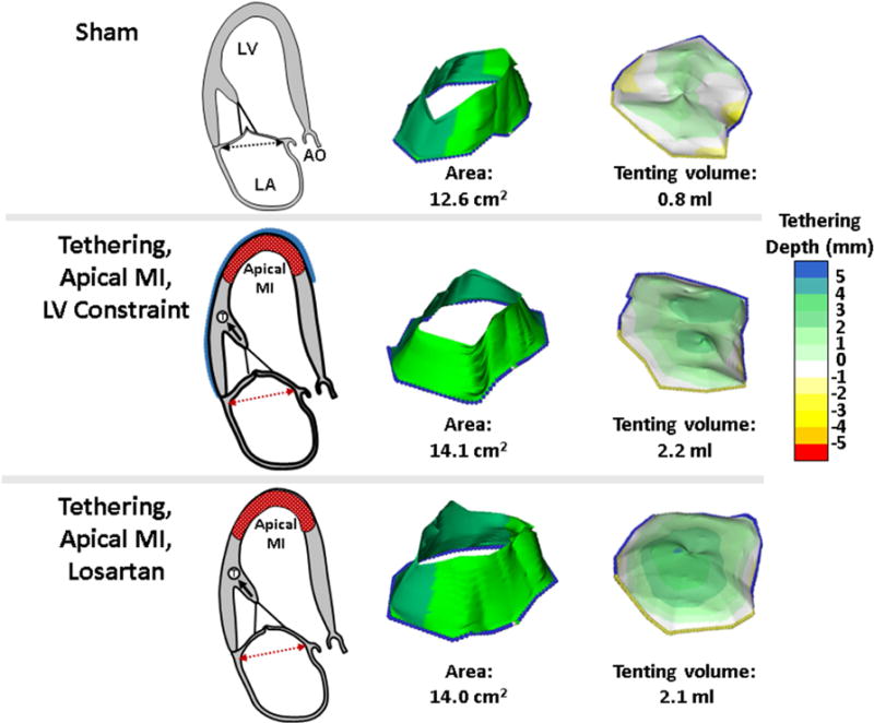
Figure 1. Model and Examples of MV Leaflet Changes.

Both the losartan-treated and untreated sheep with LV constraint display mild LV remodeling and tethering of the MV leaflets relative to the annulus with resulting increased tenting volume relative to sham-operated controls (right panels). Post-MI leaflet growth over time results in comparably increased leaflet areas (middle panels). The model provides a controlled in vivo environment with standardized tethering and apical MI that is not directly adjacent to the subvalvular apparatus. Ao = ascending aorta; LA = left atrium; LV = left ventricle, MI = myocardial infarction; MV = mitral valve.
