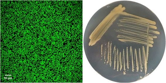
Fig. 1.

Photomicrographs of source organism. Image of P. corrugata RM1-1-4 cells using confocal laser scanning microscopy (CLSM, left) and the appearance of colony morphology after 48 h growing on NB agar medium at 25 °C (right). Image was obtained using acridin orange (0.4 mg mL−1 water) stained RM1-1-4 cells with 40× magnification. Cells under Leica TCS SP CLSM (Leica Microsystems, Wetzlar, Germany) captured and analysed using Leica Application Suite Advanced Fluorescence (LAS AF) software Version 3.5
