
Fig. 3.
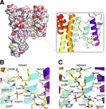
Predicted interaction mode of the putative 14–3-3γ binding site of Best1. a 14–3-3γ in complex with a phospho-peptide of Best1. Closed-up view of binding motif of hBest1 (b) and mBest1 (c). 14–3-3γ (PDB: 3UZD) recognize the putative binding motif of hBest1 and mBest1. Molecular surface of 14–3-3γ represented with charge of residues. The residues of 14–3-3γ involved in the binding are shown as white sticks. The segment of hBest1 and mBest1 display with yellow-colored stick. The secondary structure of 14–3-3γ are depicted with cartoon