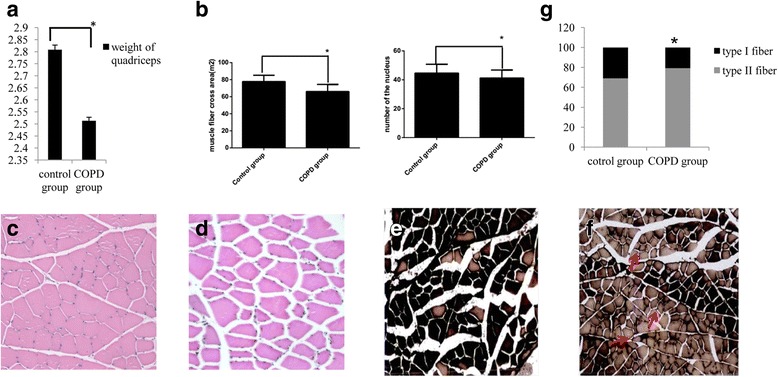
Fig. 2.

Alterations in the weight, cross-sectional area and fiber-type of the quadriceps in rats with COPD. Relative to controls, the a weight and b muscle fiber cross-sectional area and nuclear cell number of the quadriceps muscle in COPD rats were significantly reduced. n = 10, *P < 0.05,versus the control group. c Hematoxylin and eosin staining of quadriceps samples from the control group identified neat and tight muscle fibers, a narrow gap between adjacent muscle fibers, normal structures, a normal nucleus number and an even distribution (magnification, ×200). d Hematoxylin and eosin staining of quadriceps samples from the COPD group identified muscle fiber atrophy, disordered fiber arrangement, a wide gap between adjacent muscle fibers, a reduced number of nuclei and an uneven distribution of fiber sizes (indicated by black arrows; magnification, ×200) e ATPase histochemical staining (calcium-cobalt method) of quadriceps samples from the control group (magnification, ×100). Following staining, type I muscle fibers appear lighter in color than type II fibers. Control samples exhibited a larger proportion of type I muscle fibers. f ATPase histochemical staining of quadriceps samples from the COPD group (magnification, ×100). Type II fibers comprised a larger proportion of the total fibers in COPD samples (indicated by red arrows). g Quantification of muscle fiber proportions. Type I fibers were significantly decreased and type II fibers were significantly increased in the COPD group. n = 10, *P < 0.05,versus the control group. COPD, chronic obstructive pulmonary disease
