Figure 2.
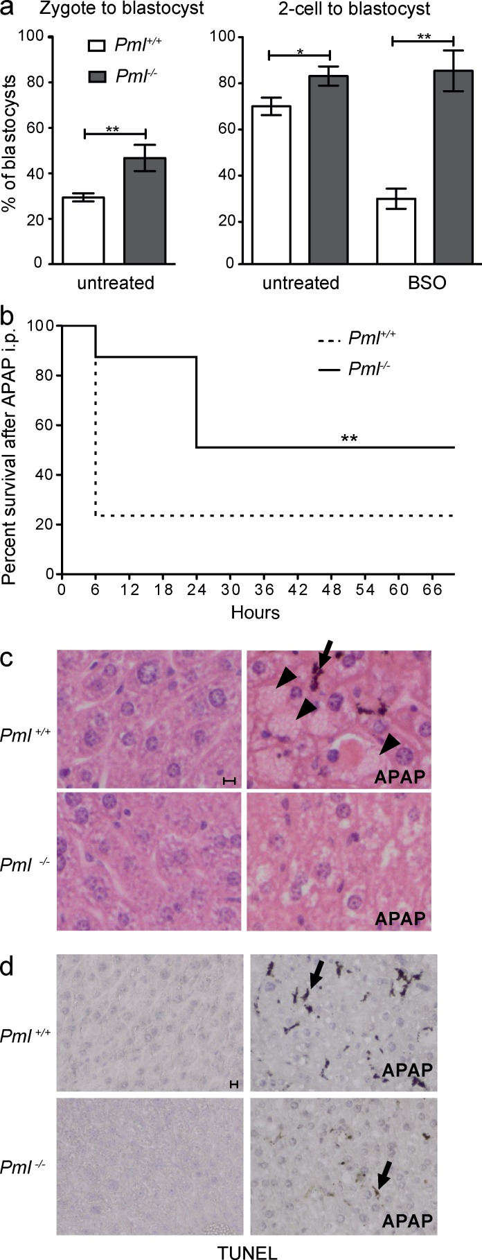
Pml−/− embryos and mice are resistant to oxidative stress. (a) Graphs representing percentage of blastocysts derived from Pml+/+ and Pml−/− preimplantation embryos. Embryos were obtained from n = 4 (untreated) and n = 3 (BSO) independent experiments involving 15–45 embryos each; error bars represent SEM (Fisher’s exact test: *, P < 0.05; **, P < 0.01). The total number of embryos was as follows: zygotes: Pml+/+, 123; Pml−/−, 106; 2-cell embryos untreated: Pml+/+, 108; Pml−/−, 140; 2-cell embryos BSO treated: Pml+/+, 45; Pml−/−, 65. (b) Survival (percentage) of Pml−/− and Pml+/+ mice after i.p. injection with APAP (time 0). A total of n = 4 independent experiments were performed (with n = 4–6 mice for each group). **, P < 0.01 (log-rank test or Gehan–Breslow–Wilcoxon tests). (c) Hematoxylin and eosin staining of liver sections from mice treated or not with APAP and sacrificed 6 h later. Note hemorrhages (arrowheads) and an apoptotic cell (arrow). (d) TUNEL performed on liver cryosections from c. Bars, 10 µm.

