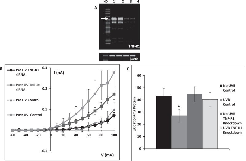
Fig. 2.
A: TNF-R1 knockdown 72 h after transfection with TNF-R1 siRNA. Lanes 1–2: control (arrow indicates TNF-R1), lanes 3–4: TNF-R1 knockdown. The lanes show protein of cells from separate wells in the same experiment. This gel is representative of 9 knockdown experiments with similar results. B: After TNF-R1 knockdown HCLE cells show diminished UVB-activated K+ currents relative to control cells following exposure to 80 mJ/cm2 UVB. (TNF-R1 data are mean ± SE of 34 cells, control data are mean of 11 cells.) C: Control cells lost 40% of intracellular K+ during a 20 min incubation following 150 mJ/cm2 UVB exposure. When TNF-R1 was knocked down, there was no loss of intracellular K+ in response to UVB. Value marked * differs significantly from all other values. Unmarked values do not differ significantly. (Mean ± SD, n = 9, ANOVA and SNK test, p < 0.05).

