
Figure 1. Direct Conversion of Young and Old Primary Adult Human Fibroblasts into Neurons via miRNA Overexpression.
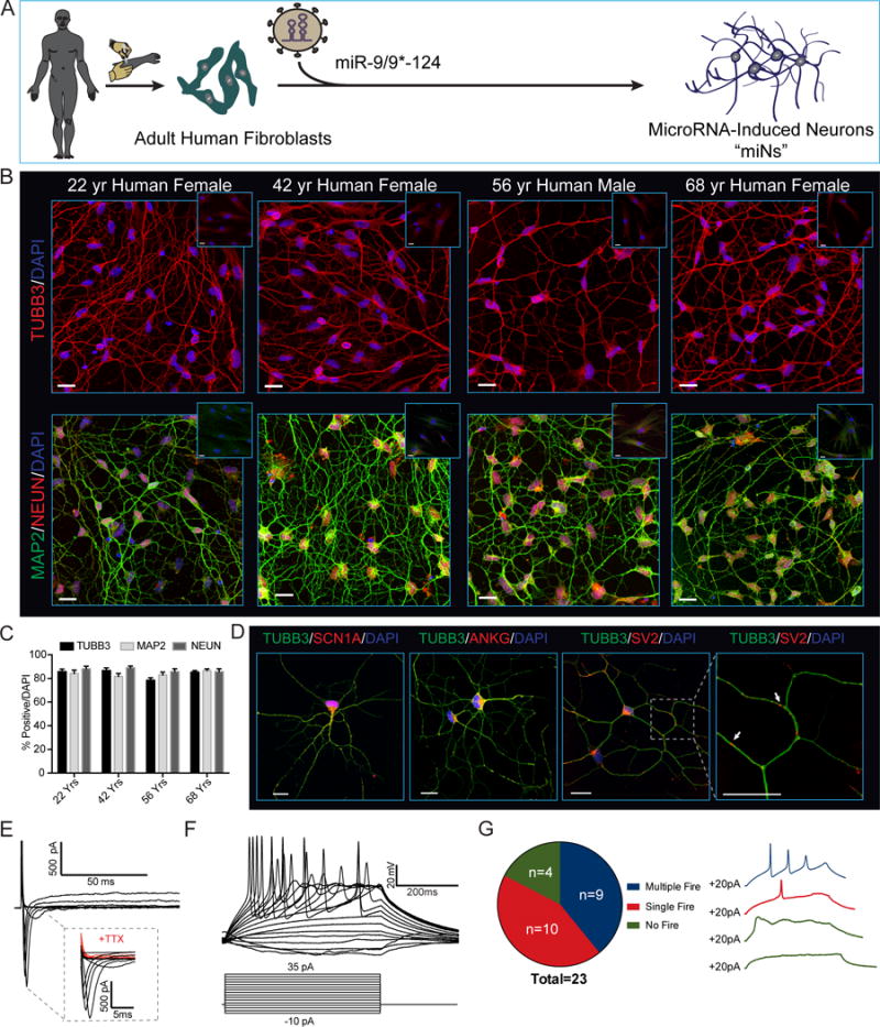
(A) Experimental scheme for miR-9/9*-124 mediated direct neuronal conversion.
(B) Adult human fibroblasts ectopically expressing miR-9/9*-124 for 35 days immunostained for the pan-neuronal markers TUBB3, MAP2 and NEUN. Insets represent starting fibroblasts co-stained as negative controls. Scale bars = 20μm.
(C) Quantification of TUBB3, MAP2 and NEUN positive cells over total number of cells (DAPI). For TUBB3 and MAP2, only cells with processes at least three times the length of the soma were counted. For NEUN, only cells with proper nuclear localization were counted. Data are represented as mean ± SEM. 22 Yr Female n = 238 cells, 42 Yr Female, n = 100 cells, 56 Yr Male n = 171 cells, and 68 Yr Female n = 216.
(D) Converted neurons display hallmark sodium channel (SCN1A), axonal initial segment (ANKG) (left) and synaptic vesicle (SV2) (right) staining patterns. Scale bars = 20μm.
(E) Representative traces of TTX-sensitive inward and potassium whole-cell currents.
(F) Repetitive AP waveforms in response to 500 ms current injections recorded from converted neurons in monoculture.
(G) Summary of AP firing patterns observed in 23 neurons recorded in current-clamp mode (left) and representative waveforms within each firing pattern recorded (right).
See also Figure S1.