
Figure 6. MiRNA-Induced Neuronal Competence Enables Motor Neuron Transcription Factors, ISL1 and LHX3, to Determine Motor Neuron Identity.
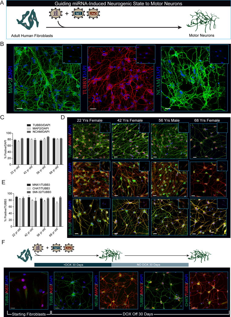
(A) Schematic of neuronal induction paradigm using miR-9/9*-124 plus ISL1 and LHX3.
(B) Representative immunohistochemistry for pan-neuronal markers in neurons generated from fibroblasts through 35 days of miR-9/9*-124, ISL1, and LHX3 co-expression. Fibroblasts were isolated from a 22-year-old female donor. Scale bars = 20μm.
(C) Quantification of 4 independent primary human fibroblast samples from male and female donors stained with TUBB3, MAP2 and NCAM. Percentages represent total number of positive cells over all cells (DAPI) and are represented as mean ± SEM. Cells (N) analyzed: 22 yr old N=TUBB3 325, MAP2 219, NCAM 275; 42 yr old N=TUBB3 304, MAP2 236, NCAM 129; 56 yr old N=TUBB3 275, MAP2 279, NCAM 213; 68 yr old N=TUBB3 282, MAP2 234, NCAM 190.
(D) Expression and correct localization of motor neuron markers in neurons converted by miR-9/9*-124 and ISL1/LHX3 as demonstrated by immunohistochemistry. MNX1, (top), CHAT (middle) and SMI-32 (bottom). Scale bars = 20μm.
(E) Quantification of (D) represents the total percentage of MNX1, CHAT and SMI-32-positive cells over TUBB3-positive cells. Data are represented as mean ± SEM. Cells analyzed: 22 yr old N=MNX1 256, CHAT 256, SMI-32 113; 42 yr old N= MNX1 151, CHAT 151, SMI-32 283; 56 yr old N= MNX1 207, CHAT 207, SMI-32 174; 68 yr old N= MNX1 151, CHAT 151, SMI-32 96.
(F) After 30 days of neuronal conversion by ectopic miR-9/9*-124 expression, Dox was removed and cells were cultured for an additional 30 days. Immunocytochemistry showing motor neurons produced by miR-9/9*-124 plus ISL1 and LHX3 (Moto-miNs) remain Ki-67 negative (2nd panel), retain expression and localization of the neuronal proteins TUBB3, NEUN, and MAP2 (2nd and 3rd panel), and express the motor neuron proteins MNX1 and CHAT (4th and 5th panel). Scale bars = 20μm.
See also Figure S5.