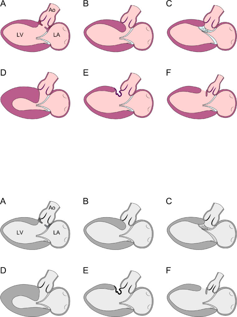
FIGURE 1.
Diagrams representing parasternal long axis transthoracic echocardiogram images depicting various anatomic types of subaortic stenosis. The figure is modified from Freedom RM. The natural and modified history of congenital heart disease. Elmsford, N.Y.: Blackwell Pub./Futura; 2004. Page 175. Figure 14C-1,1 used with permission.
A: Discrete fibrous membrane
B: Fibromuscular tunnel
C: Mitral valve tissue or its tension apparatus attached to the septum
D: Hypertrophic obstructive cardiomyopathy
E. Tissue derived from the membranous septum or tricuspid valve herniating into the LVOT through a VSD
F. Posterior displacement of the infundibular septum.
Ao: aorta; LA: left atrium; LV: left ventricle.

