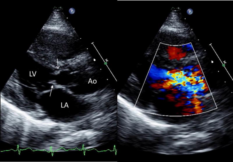
FIGURE 2.
Parasternal long-axis transthoracic echocardiographic images during systole, 2-dimensional (left) showing a discrete subaortic obstruction (top arrow, open head) approximately 1cm below the aortic valve. There is extension onto the anterior mitral leaflet (arrow with solid head). The addition of color Doppler (right) demonstrates flow acceleration with turbulence beginning at the level of the subaortic ridge.
Ao: aorta; LA: left atrium; LV: left ventricle.

