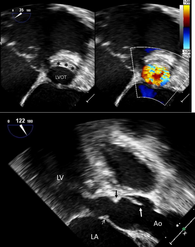
FIGURE 3.
Transesophageal echocardiography performed directly prior to surgical repair of discrete subaortic stenosis. In this example, the obstructive ridge inserts directly below the aortic valve and extends onto the anterior mitral valve.
A (Top): Long-axis view. The large white arrow identifies the aortic valve cusp, while the black arrow identifies the subaortic ridge directly below the insertion of the aortic valve. The small white arrow indicates extension of the obstructive tissue onto the anterior mitral valve leaflet. There is also hypertrophy of the basal ventricular septum proximal to the discrete subaortic membrane.
B (Bottom): Short-axis view. The black asterisks designate the subaortic ridge in cross-section.
Ao: aorta; LA: left atrium; LV: left ventricle; LVOT: left ventricular outflow tract

