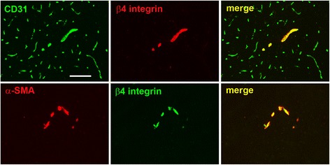
Fig. 1.

Characterization of β4 integrin expression on blood vessels in the normal brain. Top panel shows dual-IF on frozen sections of the medulla oblongata from adult mice using antibodies specific for the endothelial marker CD31 (AlexaFluor-488, green) and β4 integrin (Cy3, red). Lower panel shows dual-IF using antibodies specific for smooth muscle cell marker α-SMA (Cy3, red) and β4 integrin (AlexaFluor-488, green). Scale bar = 100 μm. Note that β4 integrin was expressed by only a fraction of CD31-positive vessels, but co-localized strongly with α-SMA
