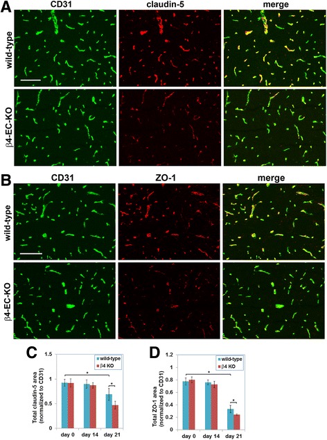
Fig. 6.

Evaluating the impact of genetic deletion of endothelial β4 integrin on endothelial tight junction protein expression in EAE. a, b Frozen sections of the medulla oblongata taken from the β4-EC-KO and WT littermate control mice at the acute stage of EAE (day 21) were dual-stained using antibodies specific for the endothelial marker CD31 (AlexaFluor-488, green) and claudin-5 (Cy3, red) or CD31 (AlexaFluor-488, green) and ZO-1 (Cy3, red) in A and B, respectively. Scale bar = 100 μm. c, d Quantification of endothelial expression of claudin-5 (c) and ZO-1 (d) in the β4-EC-KO vs. WT littermate mice. Data points represent the mean ± SEM of events observed in the medulla oblongata (n = 4 mice). Note that in the WT mice, endothelial levels of claudin-5 and ZO-1 were markedly reduced in the acute phase of EAE (day 21), and at this phase of disease, expression levels in the β4-EC-KO mice were significantly reduced compared to their WT littermates
