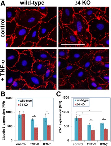
Fig. 7.

Evaluating endothelial tight junction protein expression in primary brain endothelial cells (BECs). a BECs from the β4-EC-KO and WT littermate control mice were cultured on laminin in the presence or absence of TNF-α, and claudin-5 expression was examined by IF. b, c Quantification of claudin-5 (b) or ZO-1 (c) expression by β4 integrin null and WT BECs under pro-inflammatory conditions (treatment with TNF-α or IFN-γ). All points represent the mean ± SEM of the mean fluorescent intensity (MFI) of three separate experiments. Note that while ZO-1 expression in WT BECs was significantly reduced by TNF-α and IFN-γ, expression of claudin-5 was not significantly affected by either of these cytokines. Furthermore, compared to WT BECs, while β4 integrin KO cells expressed equivalent levels of claudin-5 and ZO-1 under basal conditions, after exposure to TNF-α or IFN−γ, β4 integrin KO BECs expressed significantly lower levels of claudin-5 and ZO-1. *p < 0.05
