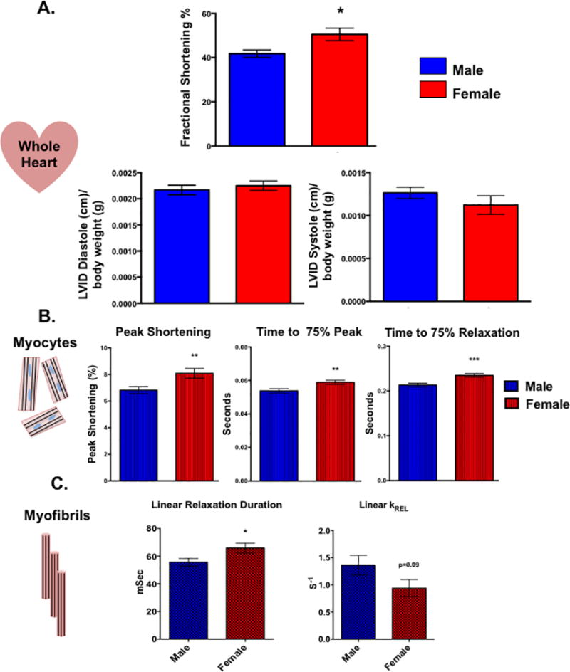
Figure 1. Cardiac function is sexually dimorphic from the level of the whole heart down to the myofibril.

(A) In vivo whole heart function as measured by echocardiography in male and female rats. *p<0.05 relative to male, N=5 female and 6 males. (B) Contractile function of electrically paced ventricular myocytes from rats of both sex. **p<0.01, ***p<0.001 relative to male, N=97 female cells and 110 male cells pooled from 6 animals of each sex. (C) Kinetic properties of left ventricular myofibrils isolated from male and female rats. Linear relaxation duration: *p<0.05 relative to female, N=21 female and 25 male myofibrils from 3 separate animals. Linear KREL: N= 15 female and 14 myofibrils from 3 separate animals. All data reported as mean ± SEM.
