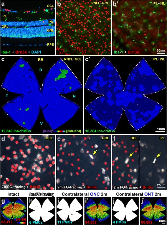
Fig. 1.

Microglial cells are homogeneously distributed in intact retinas. a Cross section showing the location of Iba1+MCs (green) and Brn3a+RGCs (red) in intact retinas. Note that some MCs in the IPL are lying on top of the INL. b Magnification from an intact flat-mounted retina focused on the RNFL/GCL showing Iba1+MCs and Brn3a+RGCs. b’ Iba1+MCs and displaced Brn3a+RGCs in the same area as b but changing the focus to the IPL/INL. c Neighbor map of an intact retina showing the distribution of MCs in the GCL. c’ Neighbor map from the same retina showing the distribution of MCs in the IPL. d Magnification from a control retina 7 days after fluorogold tracing (white), immunodetected with Brn3a (red). In these retinas, no PMCs were observed. In control retinas analyzed 2 months after tracing (e, f) some PMCs (yellow arrows) were observed in the GCL (e, f left) and IPL (f right). g Neighbor map of an intact retina showing the distribution of Brn3a+RGCs. h Retinal outline showing the position of the few PMCs found in control retinas analyzed 2 months after tracing. i, j Retinal outlines showing the location of the PMCs found in contralateral to the lesion retinas analyzed 2 months after the unilateral ONC (i) or ONT (j). i’, j’ Brn3a+RGCs neighbor maps from the same retinas as in i, j. For this and the subsequent figures: The number of cells represented is shown below each map. Neighbor maps represent the number of neighbors around each cell (dot) within a given radius and color scale. For MC representation, the radius is 0.276 mm and the scale (c, bottom) goes from 0 to 24 (purple) to ≥ 250–274 neighbors (bright green). For RGCs, the radius is 0.0552 mm and the color scale (g, bottom) goes from 0 to 4 (purple) to ≥ 35–39 (dark red) neighbors. D dorsal, V ventral, T temporal, N nasal, RR right retina, RNFL retinal nerve fiber layer, GCL ganglion cell layer, IPL inner plexiform layer, INL inner nuclear layer, OPL outer plexiform layer, ONL outer nuclear layer, ONC optic nerve crush, ONT optic nerve transection, MC microglial cell, PMC phagocytic microglial cell, m months, d days
