Fig. 3.

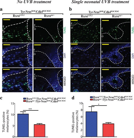
Melanomas from trigenic RXRα ep−/−|TyrNRAS Q61K|Cdk4 R24C/R24C mice display reduced apoptosis relative to their controls with functional RXRα. a, b TUNEL assay to label apoptotic cells. Apoptotic cells are indicated by green staining (top panel), blue color corresponds to DAPI staining of the nuclei (middle panel) and merged TUNEL and DAPI cells (lower panel). Overall, reduced TUNEL positive staining was observed in lesions from trigenic Rxrαep−/− mice compared to their respective controls in mice with (a) no UVB treatment as well as the (b) acute UVB treated mice. c, d Bar-graph represents TUNEL+ melanocytes/ field in no UVB and acute UVB treatment respectively
