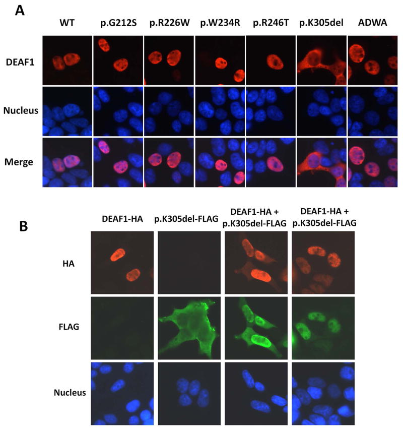
Figure 4. Subcellular localization of DEAF1 variants and changes in localization of WT DEAF1 and p.K305del in cells co-expressing both proteins.
A. Immunofluorescence analysis of WT DEAF1 and DEAF1 variants in transfected HEK293T cells revealed nuclear localization for variants p.G212S, p.R226W, p.W234R and p.R246T, and cytoplasmic localization for p.K305del. DEAF1 protein is shown in red in the upper panels, nuclei in blue in the middle panels, and merged signals in the bottom set of panels. B. Protein interactions between WT DEAF1 and p.K305del were monitored by immunofluorescence analysis. Co-expression of these two proteins resulted in nuclear and cytoplasmic localization of both proteins (columns 3 and 4). HA epitope-tagged DEAF1 protein is shown in red in the upper panel, FLAG epitope-tagged p.K305del in green in the middle, and nuclei in blue in the bottom.

