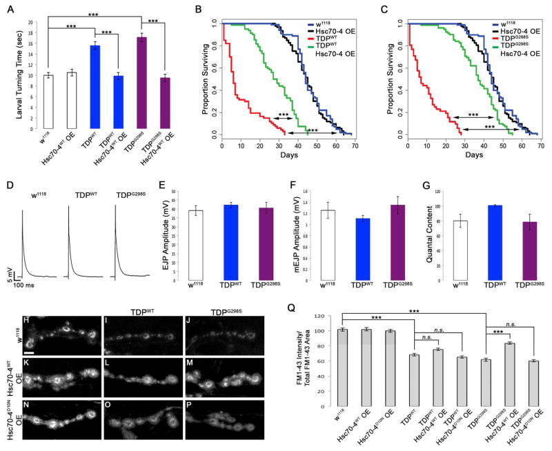
Figure 3. TDP-43 expression results in defects in SV endocytosis that are suppressed by Hsc70-4 in a variant dependent manner.
(A) Motor neuron expression of TDP-43WT or TDP-43G298S results in increased larval turning time, which is mitigated by OE of Hsc70-4. (B–C) Motor neuron expression of TDP-43 variants WT (B) or G298S (C) leads to reduced lifespan. OE of Hsc70-4 increases lifespan for both TDP-43WT (B) and TDP-43G298S (C). (D) Representative electrophysiology traces of EJPs. Genotypes indicated on top. (E–G) EJP amplitude (E), mEJP amplitude (F), and quantal content (G) measurements from electrophysiology recordings. Genotypes indicated on bottom. (H–P) Confocal images of FM1-43 dye uptake in synaptic boutons of Drosophila larvae after 5 min of stimulation in HL-3 saline containing 90 mM KCl and 2 mM Ca2+. Genotypes indicated on top and left. (Q) Quantification of FM1-43 dye uptake normalized to total FM1-43 uptake area. Note an 18 ± 1.8% reduction in FM1-43 dye uptake for TDP-43G298S, Hsc70-4 OE compared to w1118 controls (p<0.001). Scale bar (D) 10 μm.

