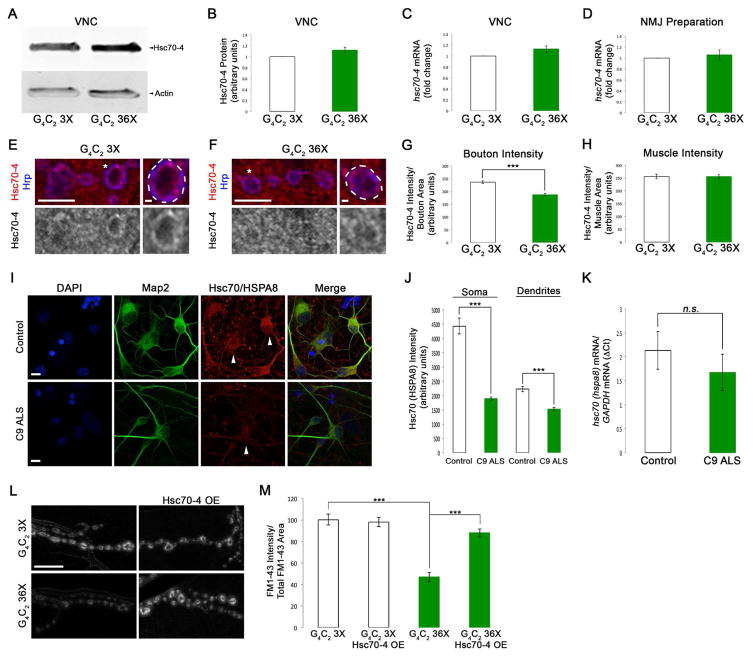
Figure 6. C9orf72 repeat expansions cause reduced Hsc70-4/HSPA8 expression and defects in SVC.
(A) WB for Hsc70-4 levels in VNCs of G4C2 expressing larvae. Genotypes indicated on bottom. Actin was used as loading control. (B) Quantification of Hsc70-4 protein levels from WBs represented as a ratio to G4C2 3X controls. (C) qPCR for hsc70-4 mRNA in VNCs from animals expressing G4C2 36X in motor neurons versus G4C2 3X controls. (D) qPCR for hsc70-4 mRNA in NMJ preparations from animals expressing G4C2 36X in motor neurons versus G4C2 3X controls. (E–F) Single confocal sections of synaptic boutons in NMJ preparations immunostained for Hsc70-4 and the neuronal membrane marker Hrp from larvae expressing G4C2 36X (F) compared to G4C2 3X controls (E). Antibodies indicated on left. (G) Quantification of Hsc70-4 intensity in synaptic boutons normalized to bouton area. (H) Quantification of Hsc70-4 intensity in muscle normalized to muscle area. (I) Confocal images of control and C9 ALS human iPSC motor neurons immunostained for DAPI, the dendritic marker Map2, and Hsc70/HSPA8. Genotypes indicated on left and antibodies indicated on top. (J) Quantification of Hsc70/HSPA8 intensity in the soma and dendrites of control and C9 ALS human iPS motor neurons. (K) qPCR for Hsc70/HSPA8 mRNA in control and C9 ALS human iPS motor neurons. (L) Confocal images of FM1-43 dye uptake at Drosophila NMJs after 5 min of stimulation in HL-3 saline containing 90 mM KCl and 2 mM Ca2+. Genotypes indicated on top and left. (M) Quantification of FM1-43 dye uptake normalized to total FM1-43 uptake area. Scale bars (E–F) 5 μm, 1 μm, (I, L) 10 μm.

