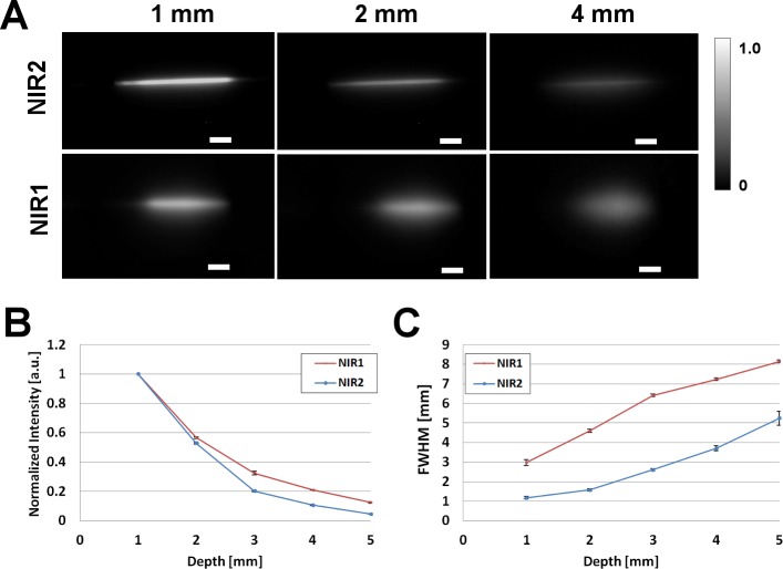
Fig 3. Intralipid® phantom study of ICG in NIR-I and NIR-II window.
(A) Fluorescence images in NIR-II (top panel) and NIR-I window of glass capillary filled with ICG in plasma (50µM) at depths of 1, 2, and 4 mm in 1% Intralipid®. 785 nm laser used for excitation. Scale bars are 50 mm. (B) Normalized intensity loss of ICG in plasma, in NIR-I and NIR-II as a function of depth. (C) Full-width-half-maximum (FWHM) of capillary glass tube filled with ICG in plasma as a function of depth in Intralipid®, showing loss of feature consistency in NIR-I compared to the NIR II. For NIR-I camera standard deviation of the signal intensity in the air was 0, which causes an indefinite value of CNR, for the NIR-I and NIR-II comparisons we plotted both Normalized Intensities and FWHM as in [24]. While for tissue phantoms and in-vivo experiments we report CNR values.

